Orthopedic Hip Surgery in Indianapolis, IN and Mooresville, IN

Hip Impingement (Labral Tears / CAM & Pincer Deformity)


There are three types of FAI: pincer, cam, and combined impingement.
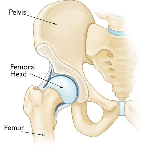
- Pincer—extra bone extends out over the normal rim of the acetabulum (socket). The labrum (cartilage seal) can be crushed under the prominent rim of the acetabulum.
- Cam—the femoral head (ball) is not round and cannot rotate smoothly inside the acetabulum (socket). A bump forms on the edge of the femoral head that grinds the cartilage inside the acetabulum.
- Combined—both the pincer and cam types are present.

Non-operative treatments:
- Activity changes—modify high impact activities and positions where your hip is maximally flexed
- Non-steroidal anti-inflammatory drugs (NSAIDs)—medications like Aleve (up to 2 pills twice a day with food) can help reduce pain and inflammation.
- Physical therapy—specific exercises can improve the range of motion of the hip, strengthen the muscles that support the joint, and alter joint mechanics so there is less impingement between the bones. Therapy can relieve some stress on the injured labrum or cartilage.
- Hip injection under fluoroscopy (x-ray)—steroid injections into the hip may also help with symptoms. Injections can also better pinpoint where hip pain may be coming from. Since the hip joint is deep, buried under many muscles, x-ray is used to make sure the medicine is delivered into the hip joint itself. This procedure is performed at the surgery center under a small amount of local anesthesia (numbing medicine at the skin).
Hip Arthroscopy


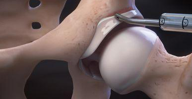
Labral repair: the “gasket” that surrounds the acetabulum (socket) is called the labrum. This provides a suction seal of the ball-and-socket joint, giving stability to the hip. If non-operative treatments have failed to relieve pain associated with a labral tear, hip arthroscopy can be used to fix the labrum and improve the stability of the joint.

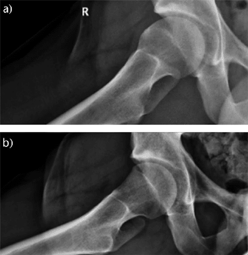
Femoroplasty: in patients who have a cam deformity (bony bump over the front of the femoral head), a burr can be used to remove the bump to prevent further pinching of the bone spur against the labrum. X-ray is used during surgery to make sure an appropriate amount of bone is being removed and at the right location. There is a small risk of fracture of the femur, so you will be on crutches for 2 weeks after this type of procedure. There is also a small risk of extra bone forming in the hip (heterotopic ossification) from the bone shavings. To prevent this, you will be prescribed a powerful anti-inflammatory called Celebrex for 2 weeks after surgery.
Acetabuloplasty: Similar to the bony bump on the femoral side, bone spurs can occur on the acetabular (socket) side. This is called a pincer deformity. The bone spur on the socket can be removed in the same manner as the bone spur on the femoral side. There is lower risk of fracture with this procedure as compared with the femoroplasty. There is still the same risk of heterotopic ossification, so patients are required to take Celebrex for 2 weeks after surgery.


Iliopsoas fractional lengthening: in a small percentage of patients, deep groin pain can be accompanied by more superficial (closer to the skin) discomfort, often caused by iliopsoas (hip flexor) tendinitis and/or bursitis. Pain can be reproduced with pushing down on a leg that is being lifted up. Some movements of the hip can also cause snapping or popping. If non-operative treatments have failed to improve pain, the iliopsoas tendon can be lengthened during hip arthroscopy. The tendon is not “cut”, but rather “lengthened” in the area where the tendon and muscle combine (musculotendinous junction) immediately in front of the hip.
